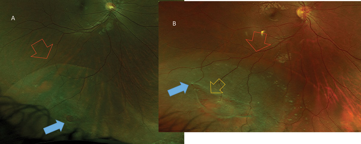
Patients with the triad of youth, myopia, and round holes in lattice degeneration deserve close observation. Young, moderate to highly myopic patients with round holes in areas of lattice degeneration seem to have a greater risk of developing this type of detachment. Surgical repair with standard scleral buckling techniques was successful in 98% of these detachments. When located inferiorly, there was a tendency for slow progression as indicated by the frequent presence of pigmented demarcation lines. Inferior detachments were slightly more common than superior detachments. The retina can begin to disintegrate over. Over 75% of the patients had refractive errors more myopic than -3 D spherical equivalent. Atrophic retinal holes are thought to be caused by these aging changes in both the retina and the vitreous humour. One of the patients were under the age of 30 years. These detachments had the following important characteristics: 1. Detachments due solely to round holes in lattice accounted for almost 2.8% of all retinal detachments treated at Wills Eye Hospital from January 1970 to August 1973. The symptoms typically resolve within 1 to 2 weeks but may not resolve fully.Round atrophic holes in lattice degeneration are an important cause of phakic retinal detachment. Some patients also experience some difficulty focusing up close. Possible side effects include an increase in pupil size in the treated eye. Side effects are reasonably uncommon, but the risk of side effects increases with the amount of lattice and treatment required. Lattice degeneration is typically treated with laser to strengthen the retina in areas where it is weak. However, other risk factors such as nearsightedness and a family history of retinal tears or detachment convey additional risks that may prompt your doctor to recommend treatment. The current literature regarding prevention of retinal detachment does not provide sufficient information to strongly support routine treatment of lesions other than symptomatic flat tears. If a new retinal tear or a retinal detachment develops, these should be promptly treated with current techniques. Among patients with lattice degeneration associated with tiny atrophic holes, only 2% will be in danger of a later retinal detachment. Retinal Hole ETIOLOGY, Atrophic retinal hole: Focal loss of retinal tissue due to retinal thinning and atrophy such as with lattice degeneration ETIOLOGY. Retinal tears are much more common and are ideally discovered and treated before they lead to a retinal detachment. retinal atrophic holes, because of massive vitreous liquid. In macular degeneration, the center of your retina begins to deteriorate. The hole may develop from abnormal traction between the retina and the vitreous, or it may follow an injury to the eye. These adhesions represent the chief clinical danger of this disease because of their ability to lead to retinal tears and detachment after PVD.ĭuring the lifetime of a patient with lattice degeneration, the likelihood of having a retinal detachment on this basis is in the range of 1% to 2%. exudative retinal detachment, macular hole induced RRD, recurrent RRD, asymptomatic RRD. A macular hole is a small defect in the center of the retina at the back of your eye (macula). Lattice degeneration is located at the edge of the retina and is associated with abnormally strong adhesions between the retina and the vitreous gel that fills the eye. Depending on symptoms and hole appearance, a decision is made whether to treat the lesion or observe. Occasionally, holes may be found in the lattice.

To discover all such lesions, it is necessary to perform a dilated eye exam combined with scleral indentation, where an instrument is used to put gentle pressure on the eyelids to better examine the edge of the retina. Lattice Degeneration represents an area of retinal thinning, usually located near the outer edge of the retina. Lattice degeneration is almost always discovered inadvertently, either in the course of a routine eye examination or in conjunction with symptoms of a PVD. Ultimately, a retinal detachment can occur following a retinal tear. Occasionally, if an acute posterior vitreous detachment (PVD) occurs, a retinal tear may be observed at a lattice lesion the tear then may produce symptoms of this event (floaters or light flashes). No symptoms are associated with this condition. What are the symptoms of Lattice Degeneration? Methods : Treatment-nave patients with atrophic holes or lattice degeneration in the retina periphery, and without visual symptoms were recruited between March. It usually shows an autosomal dominant pattern and occurs in about 8% of healthy individuals of both sexes. Lattice degeneration is a very common, inherited, congenital abnormality of the peripheral retina.


 0 kommentar(er)
